Absolute bioluminescence imaging at the single-cell level with a light signal at the Attowatt level
Bioluminescence imaging (BLI) demonstrates cellular events as a light signal at the single-cell level using a highly sensitive, cooled CCD camera. However, BLI signals are relative values and thus, images taken on different days or using different equipment cannot be compared directly. We established a reference LED light source that was characteristic of the total flux and light distribution and calibrated the BLI system as an absolute light signal. This calibrated BLI system revealed that the average light signal of beetle luciferase was at an attowatt level per sec at the single cell level.
Bioluminescent reactions have been widely used as a convenient reporter system in gene expression analysis of living cells [1–3]. Light signal from luciferin and luciferase reactions within cell populations can be measured as an average value using a luminometer. Furthermore, recent advances in bioluminescence imaging (BLI) have facilitated the demonstration of cellular events as light signals, such as the dynamics of cellular organelles or changes in promoter activity, using highly sensitive, cooled CCD cameras [4–6]. However, BLI signals are relative values and images taken on different days, using different equipment or BLI probes cannot be directly compared. For instance, light intensities cannot be compared quantitively between blue-emitting BLI and yellow-emitting BLI, as the detecting efficiencies of detectors such as CCD depend on the wavelength of the emission light. Thus, we propose absolute BLI based on a reference LED light source to overcome this challenge.

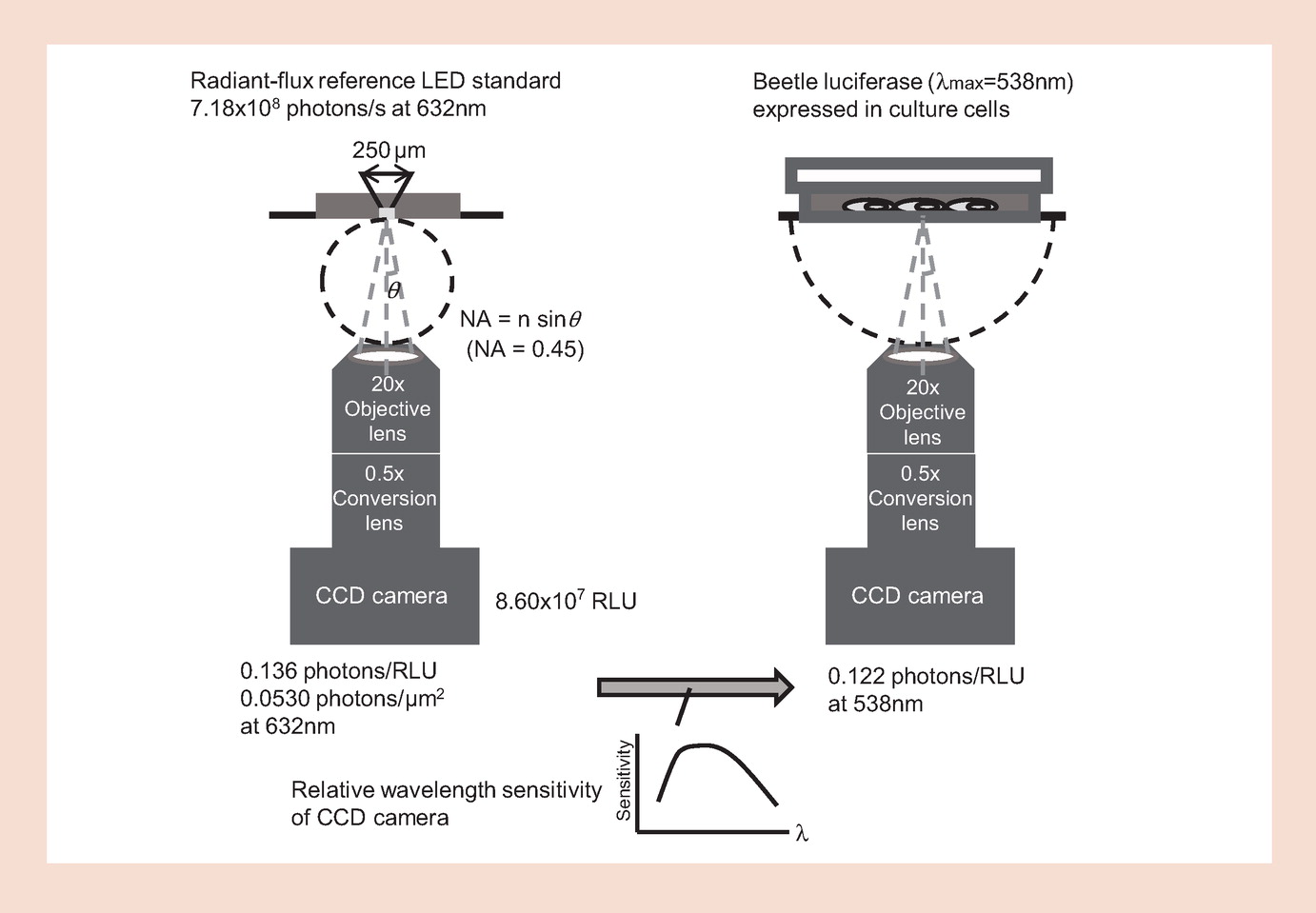
Figure 1. Illustration of the schematic of calibration for the |
For the calibration of the BLI system, we established the reference LED light source with a pulse-width modulation (PWM) control (Figure 1), which possessed a light-emitting aperture at an emission wavelength of 632 nm because of light-output stability over the long term. This source had a hole 250 μm in diameter, with total radiant flux (Rf) of 903 (±10%) pW. Total Rf (W) of the LED was evaluated by NMIJ/AIST (Tokyo, Japan) based on the absolute sphere method. The characteristics of light distribution of the source were measured using a previously described method [7]. Photon energy of the light source was found to be 3.14 × 10⁻¹⁹ J and the number of photons was 7.18 × 10⁸ per sec.
Для продолжения чтения вы можете скачать полную версию материала по ссылке ниже